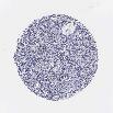
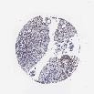
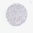
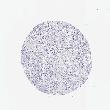
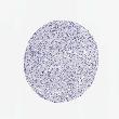
The cancer tissue page shows antibody staining in 20 different cancers. The overall cancer tissue staining statistics shows the fraction of patient samples with high, medium, low or not detected (as described by the color-coding scale in the box to the right), using all the available antibodies to the protein targets encoded by this gene. The assay and annotation is described
here.
The cancers can be ordered histologically or alphabetically.
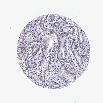
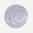
For each cancer, the staining for each available antibody is reported as the fraction of samples with antibody staining level high, medium, low or not detected (as described by the color-coding scale in the box to the right). The length of the bar represents the number of patient samples analyzed (max=12 patients).
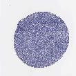
By clicking on a cancer tissue, the detailed staining data for that cancer is available, including annotated images.
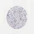
At the bottom of this page, a summary for the cancer staining for each antibody is given, together with the
literature conformity for the staining pattern revealed by that specific antibody.